When I look at the terms echocardiogram and ECG, I understand why many people assume they are simply two names for the same heart test. Both are commonly used to investigate heart problems, both are noninvasive, and both can provide important information about how the heart is functioning. However, they examine very different aspects of the heart.
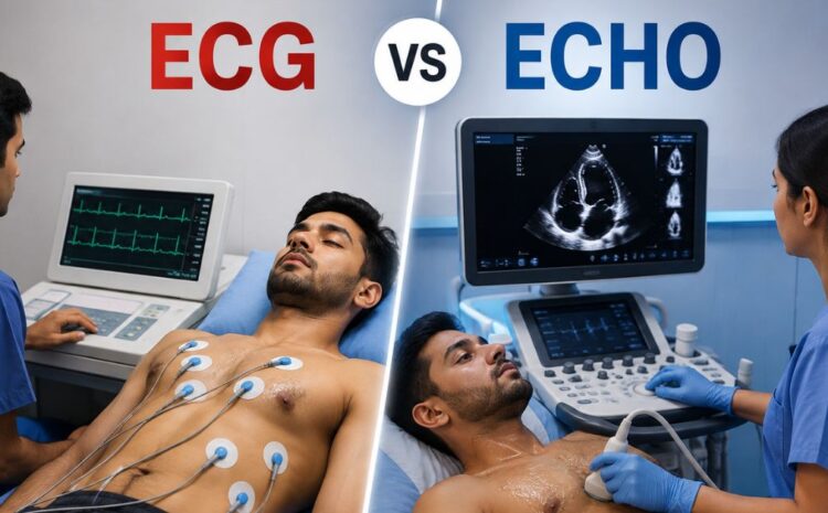
In my analysis, the simplest way to understand the difference is this: an ECG primarily records the heart’s electrical activity, while an echocardiogram uses sound waves to create moving images of the heart. The American Heart Association describes an ECG as a test that measures the electrical waves of the heart, whereas an echocardiogram uses sound waves to create pictures of the heart.
That distinction matters because the heart has both an electrical system and a physical structure. The electrical system controls the timing and rhythm of contractions, while the heart muscle, chambers, valves, and blood flow determine how effectively the heart performs its pumping function.
From my perspective, neither test is automatically “better.” The right test depends on what a healthcare professional is trying to investigate. Sometimes an ECG provides the most useful initial information. In other situations, an echocardiogram is more appropriate. There are also circumstances in which both tests are performed because they answer different clinical questions.
Key Takeaways About Echocardiogram vs ECG
The most important points I want readers to remember are straightforward:
- An ECG, also called an EKG, records the electrical activity of the heart.
- An echocardiogram is an ultrasound examination that produces moving images of the heart.
- An ECG is particularly useful for evaluating heart rate, rhythm, and electrical conduction.
- An echocardiogram can show heart chambers, valves, muscle movement, pumping function, and blood flow.
- An ECG is generally very quick and usually takes only a short time to perform.
- A standard echocardiogram commonly takes longer because the healthcare professional must obtain multiple ultrasound views.
- Both tests are generally noninvasive.
- A normal ECG does not necessarily mean that the heart’s structure and valves are normal.
- A normal echocardiogram does not necessarily rule out an electrical rhythm problem.
- Doctors may use both tests when they need information about the heart’s electrical activity and physical structure.
We can see why comparing echocardiogram vs ECG as if one replaces the other can be misleading. Their purposes overlap in some areas, but their core functions are different. Mayo Clinic specifically states that an echocardiogram uses ultrasound to create pictures of the heart and blood flow, while an ECG records the heart’s electrical activity.
What Is an ECG and What Does It Measure?
An electrocardiogram, commonly abbreviated as ECG or EKG, is a test that records the electrical signals produced by the heart. Small electrodes are placed on the skin, usually on the chest and sometimes on the arms or legs. Wires connect those electrodes to an ECG machine, which displays or records the electrical patterns.
I think of an ECG as a snapshot of the heart’s electrical behavior at a particular moment. It does not create an ultrasound picture of the heart. Instead, it translates electrical activity into recognizable waveforms that healthcare professionals can interpret.
The information can include heart rate, rhythm, and the timing and strength of electrical signals. According to the American Heart Association, an ECG can help identify conditions such as arrhythmias, heart attack, cardiomyopathy, congenital heart defects, coronary artery disease, heart failure, and some valve-related conditions.
Why Doctors Order an ECG
An ECG can be useful when symptoms suggest a possible rhythm or electrical problem. Examples include palpitations, a fast or slow heartbeat, dizziness, chest discomfort, shortness of breath, weakness, or reduced exercise tolerance. Mayo Clinic lists several of these symptoms among reasons a healthcare professional may recommend an ECG.
The test can also be important when a healthcare professional suspects that someone has experienced a heart attack. ECG patterns can provide information about current or previous heart damage and can also offer clues about reduced blood flow to the heart.
A practical example helps illustrate the distinction. Suppose a person repeatedly feels that their heart suddenly races or “flutters.” An ECG can record the electrical pattern during the test and may reveal an abnormal rhythm. If the symptoms occur only occasionally, a healthcare professional might instead consider longer-term monitoring, such as a Holter monitor or another ambulatory cardiac monitor.
What an ECG Does Not Directly Show
One common misconception I encounter in explanations of heart testing is the assumption that an ECG provides a detailed picture of the heart’s anatomy. It does not.
An ECG can provide clues about some structural changes, but its primary purpose is to record electrical activity. It does not function like an ultrasound camera that directly displays the heart chambers and valves.
For example, if the question is whether a heart valve is narrowed or leaking, an echocardiogram is generally much more directly suited to that question because it can visualize the valve and assess blood flow. Mayo Clinic identifies echocardiography as an important test for evaluating valve disease.
What Is an Echocardiogram and What Does It Measure?
An echocardiogram, often called an echo, is an ultrasound examination of the heart. It uses sound waves to create moving images that allow healthcare professionals to examine the heart’s structure and function.
In my view, the easiest mental picture is to think of an echocardiogram as a real-time ultrasound of the heart. It can provide information about the heart chambers, heart muscle, valves, and blood flow.
A standard transthoracic echocardiogram, or TTE, is performed by placing an ultrasound transducer against the chest. The transducer sends sound waves into the body and receives returning echoes. A computer converts those signals into images displayed on a monitor.
Why Doctors Order an Echocardiogram
An echocardiogram may be recommended when a healthcare professional needs information about the heart’s structure or pumping function. It can help evaluate heart failure, valve disease, heart muscle damage, murmurs, congenital heart defects, and other conditions.
The British Heart Foundation explains that an echo can examine heart structures such as valves, blood flow through major vessels, and how well the heart is pumping.
Consider a hypothetical example. A person has a heart murmur detected during a physical examination. The central question may be whether a valve abnormality is responsible for the sound. In that situation, an echocardiogram can provide much more direct structural information than a standard ECG.
Another hypothetical example involves suspected reduced pumping ability. An echocardiogram can provide information about how well the heart muscle contracts and how blood moves through the chambers and valves. That makes it particularly valuable when the concern involves mechanical pumping rather than an isolated electrical rhythm problem.
Echocardiogram vs ECG: The Main Differences
When I compare echocardiogram vs ECG, I focus first on what each test actually measures. The distinction becomes much easier once that point is clear.
| Feature | ECG | Echocardiogram |
|---|---|---|
| Full name | Electrocardiogram | Echocardiogram / echocardiography |
| Main technology | Electrical sensors and recording | Ultrasound |
| Primary purpose | Records electrical activity | Produces images of heart structure and motion |
| Main information | Heart rate, rhythm, electrical conduction | Chambers, valves, muscle movement, pumping, blood flow |
| Typical procedure | Electrodes placed on skin | Ultrasound transducer moved over chest for standard TTE |
| Produces moving heart images? | No | Yes |
| Useful for rhythm problems? | Yes | Not its primary purpose |
| Useful for valve structure and function? | Limited clues | Yes |
| Radiation | No | No |
| Commonly noninvasive? | Yes | Yes |
| Can be used with other cardiac tests? | Yes | Yes |
The most important takeaway from this comparison is that the two tests answer different questions. If the question is “What is my heart’s electrical rhythm doing?” an ECG is highly relevant. If the question is “What does my heart look like, how is it moving, and how are the valves and blood flow functioning?” an echocardiogram is more directly relevant.
Neither test should be viewed as a universal replacement for the other.
How an ECG Is Performed
An ECG is generally a simple and quick procedure. The person typically lies still while electrodes are placed on the skin. The electrodes detect the heart’s electrical signals, and the machine records them.
The American Heart Association emphasizes that an ECG does not send electricity into the body; it only records electrical activity.
A practical scenario might involve someone experiencing unexplained palpitations. A healthcare professional may perform an ECG while the symptoms are occurring. If the abnormal rhythm is present during the recording, the ECG may provide useful evidence about its type.
The limitation is that a standard ECG records only a relatively brief period. If an intermittent rhythm problem does not occur during the test, the result may not capture it. In those circumstances, longer monitoring may be considered. Mayo Clinic describes Holter and event monitoring as options for recording heart activity over longer periods.
Does an ECG Hurt?
In general, an ECG is painless. The electrodes are attached to the skin, and the machine records signals without delivering electrical current into the body. The American Heart Association describes the test as having no pain or risk from the ECG recording itself, although removing adhesive electrodes can sometimes cause minor skin discomfort.
How an Echocardiogram Is Performed
For a standard transthoracic echocardiogram, a healthcare professional applies gel to an ultrasound transducer and moves the transducer across different areas of the chest. The device sends sound waves toward the heart and receives the echoes that return. A computer processes that information into moving images.
The person may be asked to change position or breathe in a particular way so that better images can be obtained. The healthcare professional may capture multiple views of the chambers, valves, walls, and blood flow.
An ECG may sometimes be performed at the same time. This is not because the tests are identical, but because the additional electrical information can complement the structural information obtained from the echocardiogram. Mayo Clinic notes that an ECG is often performed during an echocardiogram examination.
Different Types of Echocardiograms
Not every echocardiogram is performed in exactly the same way. The appropriate type depends on the clinical question.
A transthoracic echocardiogram is the standard form and obtains images through the chest wall. A transesophageal echocardiogram uses a specialized probe positioned in the esophagus to obtain detailed views from a different angle. Stress echocardiography evaluates the heart before and after exercise or medication that makes the heart work harder.
This variety is important because the phrase “echocardiogram” can refer to several related examinations. The preparation, duration, and experience can differ depending on which type is required.
What Can Each Test Reveal?
The easiest way I explain the difference is to match each test with the type of information it provides.
| Clinical Question | ECG | Echocardiogram |
| Is the heart rhythm abnormal? | Strongly useful | Not the primary test |
| Is the heart beating unusually fast or slow? | Yes | Can show heart activity but is not primarily a rhythm test |
| Are there signs suggesting a heart attack? | Can provide important clues | Can assess heart muscle and function |
| Are heart valves narrowed or leaking? | Limited information | Strongly useful |
| How well is the heart pumping? | Provides indirect clues | Directly assesses structure and motion |
| What is the size and shape of heart chambers? | May provide indirect clues | Directly visualizes them |
| How is blood moving through valves and chambers? | No direct blood-flow imaging | Yes |
| Is there a structural abnormality? | May provide clues | Direct visualization is possible |
| Is intermittent rhythm monitoring needed? | ECG-based monitoring can help | Not its primary role |
| Is a quick electrical snapshot needed? | Yes | No |
I would not interpret this table as meaning that one test is always superior. Instead, it shows why healthcare professionals choose tests based on the question they need answered.
When Doctors May Order Both an ECG and Echocardiogram
There are many situations in which the most useful approach is not choosing between the tests but using both.
For example, suppose a person has shortness of breath and a healthcare professional wants to investigate possible heart disease. An ECG may provide information about rhythm and electrical activity, while an echocardiogram can examine the heart’s structure, pumping action, valves, and blood flow. Mayo Clinic lists both tests among the diagnostic tools used in evaluating heart disease.
A second hypothetical example involves a person with a heart murmur and occasional palpitations. The murmur may lead to an echocardiogram because valve structure and blood flow need to be assessed. The palpitations may lead to an ECG or longer-term rhythm monitoring because electrical activity needs to be examined.
This is why I believe the phrase “echocardiogram vs ECG” should not always be interpreted as a competition. In actual medical evaluation, they can be complementary.
What Does a Normal ECG Mean?
A normal ECG generally means that the electrical activity recorded during the test does not show certain abnormalities that the healthcare professional is looking for. It can provide reassurance, but it should not automatically be interpreted as proof that every aspect of heart health is normal.
For instance, an ECG is not designed to provide the detailed structural images produced by an echocardiogram. A person could therefore have a normal ECG while still requiring further evaluation for a valve problem or another structural issue.
The opposite situation is also possible. An echocardiogram may show normal heart structure and pumping function while a person has an intermittent rhythm problem that was not captured during the examination.
From my perspective, this is one of the most important concepts to understand: a normal result is always interpreted in the context of the symptoms, medical history, physical examination, and other available information.
What Does a Normal Echocardiogram Mean?
A normal echocardiogram generally indicates that the structures and functions evaluated during the examination do not show significant abnormalities according to the interpreting healthcare professional.
It can provide useful information about chamber size, heart muscle movement, valve function, and blood flow. However, it does not answer every possible cardiac question.
For example, an echocardiogram is not primarily a test for detecting every type of abnormal heart rhythm. If someone experiences episodes of an irregular heartbeat, an ECG or longer-term cardiac monitoring may be more appropriate for investigating that symptom.
We can therefore think of the two tests as complementary windows into different dimensions of heart health.
What the Evidence Says About Their Different Roles
A particularly useful explanation comes from the Mayo Clinic because it clearly distinguishes the technologies involved.
“An echocardiogram uses sound waves to create pictures of the heart.”
Mayo Clinic
I consider this quotation useful because it captures the central purpose of echocardiography without unnecessary technical language. The test is fundamentally an imaging examination.
The same source makes the ECG distinction equally clear:
“An ECG or EKG records the heart’s electrical activity.”
Mayo Clinic
In my view, these two statements provide the foundation for understanding the entire comparison. One test primarily records electrical signals; the other creates images using ultrasound.
The American Heart Association also emphasizes the role of the ECG in examining the heartbeat:
“An electrocardiogram measures the electrical activity of the heartbeat.”
American Heart Association
This matters because electrical activity is what coordinates the timing of the heart’s contractions. If the electrical system is abnormal, the resulting rhythm may be too fast, too slow, irregular, or otherwise abnormal.
Common Situations and Which Test May Be More Relevant
I want to emphasize that only a qualified healthcare professional can determine which test is appropriate for an individual. Still, general examples can help explain the reasoning.
Palpitations and Suspected Arrhythmia
If the main concern is a racing, fluttering, skipped, or irregular heartbeat, an ECG is often particularly relevant because it records electrical activity.
If symptoms are intermittent, however, a brief ECG may not capture the abnormal rhythm. Longer-term ECG-based monitoring may then be considered.
Heart Murmur
When a healthcare professional hears a murmur and wants to determine whether a valve or structural problem is responsible, an echocardiogram can be especially useful.
Mayo Clinic identifies echocardiography as the main test used to determine the cause of a concerning heart murmur because it can show the beating heart, valves, and blood flow.
Suspected Heart Failure
Both tests may contribute information when heart failure is being evaluated. An ECG can reveal electrical abnormalities, while an echocardiogram can provide information about the heart’s structure and pumping function. The American Heart Association lists both ECG and echocardiography among tests used in diagnosing heart failure.
Possible Heart Attack
An ECG can be particularly important in suspected acute coronary events because it can reveal electrical patterns associated with heart attack. Other tests, including blood tests and imaging, may also be necessary depending on the situation.
An echocardiogram may provide additional information about heart muscle movement and function, but it does not replace the full diagnostic assessment.
Step-by-Step: How to Understand Which Test You Are Being Asked to Have
When a healthcare professional recommends a cardiac test, I suggest focusing on the question the test is intended to answer rather than simply memorizing the name.
Step 1: Identify the Main Symptom or Concern
Think about why the test was recommended. Is the concern an irregular heartbeat, chest discomfort, breathlessness, a murmur, reduced exercise tolerance, or a known heart condition?
The reason for testing helps determine what information is most useful.
Step 2: Ask What the Test Measures
If the answer is electrical activity, the test is likely an ECG or an ECG-based monitor.
If the answer involves images of the heart, valves, chambers, pumping action, or blood flow, an echocardiogram is likely involved.
Step 3: Find Out Whether the Test Is Standard or Specialized
A standard ECG differs from prolonged ECG monitoring. Likewise, a standard transthoracic echocardiogram differs from stress or transesophageal echocardiography.
Understanding the specific test can make the instructions much less confusing.
Step 4: Ask How the Result Will Be Used
A test result rarely stands alone. The healthcare professional may compare it with symptoms, examination findings, previous results, blood tests, imaging, or other information.
Step 5: Follow the Preparation Instructions
Preparation can vary depending on the test. A standard ECG is usually straightforward, while certain specialized echocardiograms or stress studies can require specific instructions.
For example, Mayo Clinic notes that preparation for some stress echocardiograms may include restrictions involving caffeine, depending on the circumstances and the healthcare team’s instructions.
Common Misconceptions About Echocardiogram vs ECG
One misconception is that an echocardiogram is simply a more advanced ECG. I do not think that is an accurate way to describe the relationship. They are based on different technologies and measure different forms of information.
Another misconception is that a normal ECG proves that the heart is structurally healthy. It does not provide the same direct structural assessment as an echocardiogram.
A third misconception is that an echocardiogram can detect every rhythm abnormality. It is not primarily designed for that purpose.
Some people also assume that an ECG and EKG are different tests. They are not. ECG stands for electrocardiogram, while EKG is another abbreviation for the same test. Mayo Clinic uses both terms when describing the electrical recording of the heart.
Finally, I would avoid assuming that a doctor ordering both tests means something is necessarily seriously wrong. Sometimes the tests are ordered because they provide complementary information.
Advantages and Limitations of Each Test
An ECG has the advantage of being quick, noninvasive, and highly useful for assessing electrical activity. It can provide important information about heart rhythm and can help identify patterns associated with heart attacks and other cardiac conditions.
Its main limitation is that a standard recording represents only a short period. Intermittent abnormalities may not appear during those few moments.
An echocardiogram provides a different advantage: it can show the heart in motion. It can examine structures and blood flow, making it particularly useful for valve disease, heart muscle function, chamber abnormalities, and other structural questions.
Its limitations are different. It is not primarily a test for electrical rhythm abnormalities, and the usefulness of the examination depends on obtaining adequate images and selecting the appropriate echocardiographic technique.
How Stress Testing Changes the Comparison
The comparison becomes more interesting when stress testing is involved.
A stress ECG evaluates electrical activity while the heart is working harder, commonly during exercise. A stress echocardiogram adds ultrasound imaging to examine how the heart muscle responds to increased workload.
Mayo Clinic explains that stress echocardiography can involve images before and after exercise, with ECG monitoring during the test.
This provides an excellent example of how different heart tests can work together. The ECG supplies electrical information during stress, while echocardiography supplies imaging information about the heart’s response.
I believe this also demonstrates why there is no universal answer to “Which is better?” The correct answer depends on what the healthcare team needs to learn.
Can an Echocardiogram Detect a Blockage?
This question deserves careful clarification. A standard echocardiogram does not directly show whether a coronary artery is blocked. Mayo Clinic specifically notes that a standard echocardiogram does not directly show a blockage of a heart artery. A stress echocardiogram may provide evidence suggestive of reduced blood flow by showing changes in heart muscle movement when the heart is working harder, but additional imaging may be needed to confirm a coronary blockage.
Therefore, I would not interpret an echocardiogram as a direct substitute for coronary angiography or coronary CT angiography when the clinical question specifically concerns an artery blockage.
This distinction is important because people sometimes assume that any heart imaging test can directly visualize every part of the cardiovascular system.
Can an ECG Detect Heart Problems?
Yes, an ECG can provide important information about several heart problems, but its capabilities have limits.
The test can identify abnormal heart rhythms, changes in electrical conduction, and patterns that may indicate a current or previous heart attack. It can also provide clues associated with some structural and cardiovascular conditions.
However, an ECG should not be treated as a complete structural examination of the heart.
For example, if the question involves whether a valve is leaking, an echocardiogram is generally more informative because it can visualize the valve and assess blood flow.
What Happens After the Tests?
After an ECG, the recorded electrical pattern is interpreted by a qualified healthcare professional. Depending on the situation, results may be discussed relatively quickly, although the timing can vary.
After an echocardiogram, the recorded images are reviewed and interpreted. If the images show an abnormality or if more information is needed, additional testing may be recommended. Mayo Clinic notes that further testing or referral to a cardiologist may follow depending on the results.
I think patients benefit from remembering that a test result is not the same thing as a final diagnosis in every situation. Interpretation depends on the reason for testing and the broader clinical picture.
When Should You Seek Urgent Medical Attention?
The choice between an ECG and an echocardiogram should never delay urgent medical care when severe symptoms are occurring.
Symptoms such as significant chest pain or pressure, severe shortness of breath, fainting, or other potentially serious signs require prompt medical assessment. An ECG can be an important part of evaluating suspected heart attack, but emergency evaluation may involve several tests and treatments rather than relying on one examination. The American Heart Association notes that ECGs are used in diagnosing heart attack and that other tests can also be required.
From my perspective, the practical lesson is simple: do not try to decide at home whether an ECG or echocardiogram is sufficient when serious symptoms are occurring. Seek appropriate medical evaluation.
Expert Recommendations for Understanding Your Heart Test
My first recommendation is to ask what specific clinical question the test is intended to answer. That single question can make unfamiliar terminology much easier to understand.
Second, I recommend asking whether the test is a standard examination or a specialized version. “ECG” could refer to a brief resting recording or be part of longer-term monitoring. “Echocardiogram” could refer to a standard transthoracic examination, stress echocardiography, or another specialized study.
Third, do not compare results from the two tests as though they use the same scale. An ECG report and an echocardiogram report contain different types of information.
Fourth, if a healthcare professional recommends both tests, I would not assume that the second test is unnecessary simply because the first was normal. The tests can answer different questions.
Finally, I believe the safest approach is to interpret results with the healthcare professional who knows why the test was ordered. A number or phrase on a report can have a very different meaning depending on symptoms, age, medical history, medications, and previous test results.
Echocardiogram vs ECG: Which Test Is Better for Specific Questions?
The following comparison summarizes the practical decision-making process without suggesting that readers should diagnose themselves.
| Situation or Question | Test That May Be Particularly Useful | Why |
| Suspected irregular heartbeat | ECG | Records electrical rhythm |
| Palpitations occurring during examination | ECG | May capture the rhythm during symptoms |
| Intermittent palpitations | ECG-based longer monitoring may be considered | A brief ECG may miss an intermittent event |
| Heart murmur | Echocardiogram | Examines valves, structure, and blood flow |
| Suspected valve disease | Echocardiogram | Provides direct structural and flow information |
| Assessing heart pumping function | Echocardiogram | Shows heart muscle movement and function |
| Suspected heart attack | ECG plus other appropriate tests | ECG can reveal important electrical patterns |
| Evaluating structural heart abnormalities | Echocardiogram | Provides direct heart imaging |
| Evaluating electrical conduction | ECG | Measures electrical signals |
| Assessing response to exercise | Stress ECG and/or stress echocardiogram | Different versions provide complementary information |
| Evaluating suspected heart failure | ECG and echocardiogram may both be used | They provide different types of information |
| Looking for coronary artery blockage | Depends on clinical situation; additional imaging may be needed | Standard echo does not directly visualize coronary blockages |
The key takeaway is that the “best” test is determined by the clinical question. In many cases, the most useful evaluation involves several pieces of information rather than one isolated test.
Conclusion
In my view, the central lesson from comparing an echocardiogram vs ECG is that these tests examine different aspects of heart health. An ECG records electrical activity and is especially useful for understanding heart rate, rhythm, and electrical conduction. An echocardiogram uses ultrasound to create moving images that can show heart structure, valve function, pumping action, and blood flow.
I believe neither test should be described as universally better. Their value depends on what a healthcare professional is trying to investigate. Someone with palpitations may need electrical monitoring, while someone with a murmur or suspected valve problem may benefit from echocardiography. In other situations, both tests can provide complementary information.
The practical next step is to ask why a particular test has been recommended and what question the healthcare professional wants it to answer. If you already have an ECG or echocardiogram report, I recommend discussing the findings with the clinician who ordered the examination rather than interpreting isolated measurements without context.
Frequently Asked Questions
What is the main difference between an echocardiogram and an ECG?
The main difference is what each test measures. An ECG records the heart’s electrical activity, including information about heart rate, rhythm, and electrical conduction. An echocardiogram uses ultrasound to create moving images of the heart, allowing healthcare professionals to examine structures such as chambers and valves as well as blood flow and pumping function.
Which is better, an echocardiogram or ECG?
Neither test is universally better. An ECG is generally more directly useful when the main question involves electrical activity or an abnormal rhythm. An echocardiogram is more directly useful when the concern involves heart structure, valves, blood flow, or pumping function. In some situations, a healthcare professional may order both because they provide complementary information.
Can an ECG detect a heart blockage?
An ECG can provide important clues about reduced blood flow and may show patterns associated with a heart attack, but it does not directly visualize every coronary artery blockage. Depending on the clinical situation, additional tests such as imaging or coronary angiography may be required.
Can an echocardiogram detect an irregular heartbeat?
An echocardiogram can show the heart beating during imaging and may be performed alongside ECG monitoring, but its primary purpose is to evaluate heart structure and function rather than diagnose every rhythm disorder. An ECG is the more direct test for recording electrical activity, while longer-term ECG monitoring may be considered when symptoms occur intermittently.
Is an echocardiogram painful?
A standard transthoracic echocardiogram is generally noninvasive and painless. The healthcare professional applies ultrasound gel and moves a transducer over the chest to obtain images. Some specialized echocardiograms, such as transesophageal echocardiography, involve a different procedure and may require sedation or other preparation.
Is an ECG painful?
An ECG is generally painless. Small adhesive electrodes are placed on the skin to detect electrical signals from the heart. The machine records those signals rather than sending electricity into the body. Some people may experience minor discomfort when adhesive electrodes are removed.
How long does an echocardiogram take?
The duration varies depending on the type of echocardiogram. Mayo Clinic states that most echocardiograms take less than an hour, although specialized examinations can involve additional observation or preparation.
Can you have an ECG and echocardiogram on the same day?
Yes, they can be performed during the same clinical evaluation when both types of information are needed. Mayo Clinic notes that an ECG is often performed at the same time as an echocardiogram. The reason is that the ECG records electrical activity while the echocardiogram provides ultrasound images of the heart.
Sources and References
- Mayo Clinic, “Echocardiogram.”
- Mayo Clinic, “Electrocardiogram (ECG or EKG).”
- Mayo Clinic, “Heart Disease: Diagnosis and Treatment.”
- American Heart Association, “Electrocardiogram (EKG or ECG).”
- American Heart Association, “Echocardiogram (Echo).”
- British Heart Foundation, “Echocardiogram.”
Disclaimer
This article is intended for general educational purposes and should not be considered medical advice, diagnosis, or treatment. Test selection and interpretation depend on individual symptoms, medical history, physical examination, and other clinical information. I recommend discussing any concerns about an ECG, echocardiogram, abnormal result, or heart-related symptoms with a qualified healthcare professional. If serious or potentially life-threatening symptoms occur, seek urgent medical evaluation rather than relying on information in this article.